
CBCT Imaging
CBCT Imaging
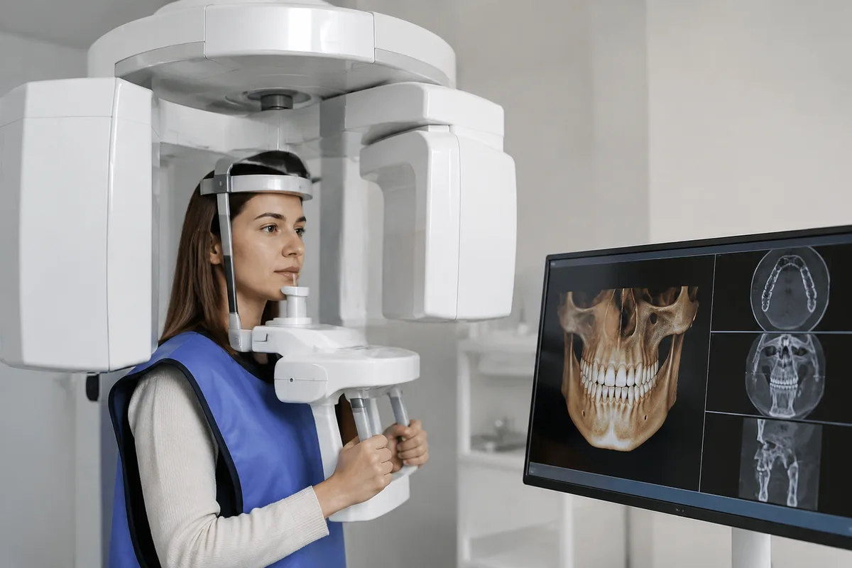
Cone-beam computed tomography, or CBCT, is a specialized imaging technology that gives a three-dimensional view of the teeth, jaws, airway, and surrounding skeletal structures. At Synera Dental Wellness, CBCT imaging is used to assess skeletal and airway structure, providing a fuller picture than a flat, two-dimensional image can offer.
What CBCT Imaging is
A CBCT scanner captures a series of images as it rotates around the head, then reconstructs those images into a detailed 3D model. Unlike a traditional dental X-ray, which produces a single flat image, CBCT allows a clinician to view bone, tooth, and airway structures from any angle and at any depth.
This technology grew out of the same principles as hospital CT scanning but was adapted specifically for dental and facial imaging, using lower radiation doses and a smaller footprint suited to a dental office. It improved on older imaging methods by capturing spatial relationships — how the jaw joints, sinuses, airway, and teeth relate to one another in three dimensions — rather than relying on a single projection that can hide or distort those relationships.
Because CBCT shows both hard tissue and airway space, it is commonly used whenever a clinician needs to understand structure, not just the surface appearance of teeth.
How we use it at Synera Dental Wellness
CBCT imaging plays a role in evaluations where skeletal and airway structure matter to diagnosis and treatment planning. Dr. Jeremy Montrose's practice is built around a physiologic, airway-focused approach to dentistry — one that considers the temporomandibular joint (TMJ), sleep and breathing patterns, and growth and development from infancy through adulthood. Understanding the underlying skeletal and airway anatomy is part of that approach, which is why CBCT evaluation appears among the services offered here.
CBCT scans can inform assessments related to TMJ evaluation, airway-focused dentistry, sleep-disordered breathing, and skeletal expansion, since each of these areas depends on a clear view of how the jaws, joints, and airway are structured. The imaging also works alongside other on-site tools: digital X-rays for routine diagnostic needs, and custom 3D-printed appliances, which are designed using precise digital scans for an accurate fit.
Where a case calls for it, a CBCT scan gives the clinical team information that a standard X-ray alone cannot provide, supporting more informed conversations about diagnosis and treatment options.
What it means for you
For patients, CBCT imaging generally means a clearer, more complete picture of what is happening beneath the surface — without requiring separate imaging visits or guesswork. Because the scan produces a 3D reconstruction, it can help clarify findings that might otherwise be ambiguous on a flat image.
- Comfort — the scan is non-invasive and does not require injections or contrast dye.
- Clarity — a 3D view can make it easier to understand and discuss findings about bone, joints, and airway space.
- Efficiency — a single scan can capture information relevant to multiple areas of evaluation at once.
These are general benefits associated with CBCT technology and should be understood as educational information rather than a guarantee of any particular result.
What to expect during your visit
Before the scan, you will be positioned so the imaging area is properly aligned; no special preparation, such as fasting, is typically required. You may be asked to remove metal objects like jewelry or glasses, since these can interfere with image quality.
During the scan, you will stand or sit while the scanner arm rotates around your head. Most CBCT scans take well under a minute of actual scanning time, though the full appointment — including positioning and any follow-up discussion — takes longer. You should feel no discomfort; the process is similar to having a panoramic dental X-ray taken.
After the scan, the images are reviewed as part of your evaluation, and findings are discussed with you as they relate to your specific concern, whether that involves the TMJ, airway, orthodontic planning, or another area of care.
Frequently asked questions
Is CBCT imaging safe?
CBCT imaging uses radiation, similar in principle to other dental X-rays, though the dose and exposure area are specific to the type of scan taken. Your clinical team will only recommend a scan when it provides information relevant to your care.
Does the scan hurt?
No. The scan involves no needles, contrast, or physical contact beyond standing still while the scanner rotates around your head. Most patients find it no different from having a panoramic X-ray taken.
How long does a CBCT scan take?
The scanning portion itself is brief, typically well under a minute. The full appointment, including positioning and any discussion of the images, will take longer depending on the reason for the scan.
Who might need a CBCT scan?
CBCT imaging is often used when a clinician needs to evaluate skeletal structure or airway space, such as during TMJ evaluations, airway-focused assessments, or orthodontic and skeletal expansion planning. It is not needed for every visit, only when a three-dimensional view of these structures is relevant to your care.
If you have questions about CBCT imaging or would like to learn whether it is relevant to your care, reach out to Synera Dental Wellness to schedule a visit.
This content is for general information only and isn't dental or medical advice. Talk with your dentist or orthodontist about your own situation. If you have a medical emergency, call 911.